
Trichoscopy over time: measuring density loss after a hair transplant
Trichoscopy can measure density and hair thickness objectively, year by year. No other method separates ongoing loss of your own hair from failing grafts as reliably.
Trichoscopy gives objective measures of density and hair shaft thickness over several years, and it's the only reliable way to tell ongoing loss of your own hair from failing grafts. Almost no clinics repeat the measurement, which is why the data is missing.
What does trichoscopy tell you that a photo doesn't?
Trichoscopy gives numbers instead of impressions. It measures how many follicular units there are per cm², the number of hairs per unit, hair shaft thickness and how much it varies (the hallmark of androgen-driven thinning), the proportion of vellus hairs and signs of inflammation around the follicles. These values change before anything shows in a photo.
A photo shows how your hair looks. Trichoscopy gives you measurable values.
- Number of follicular units per cm², counted in a field of known size, not estimated.
- Number of hairs per follicular unit. This falls as the follicles shrink.
- Hair shaft thickness, and even more importantly, how much the thickness varies between neighbouring hairs. That's the classic sign that androgens are thinning the hair.
- The proportion of vellus hairs, which rises as miniaturisation progresses.
- Findings around the follicles linked to the inflammation that is part of androgenetic alopecia.
The key point is that these values shift before the hair looks any different. An area can seem completely unchanged even though the spread of hair thickness has changed a lot.
Why should trichoscopy be repeated after a transplant?
If the result looks thinner a few years later, it may be down to grafts that failed, your own hair around the grafts continuing to disappear, or the donor area itself being affected. Each calls for completely different action. A photo won't give you the answer. Repeat measurements in the recipient area, in untreated scalp and in the donor zone will, because each site gets its own numbers.
If you notice after five or eight years that the result seems thinner, there are three possible explanations, and each needs its own solution.
- The grafts failed. Grafts that never took are already visible at twelve months. They don't gradually dwindle afterwards.
- Your own hair is disappearing. The hair between and around the grafts keeps miniaturising. This is the most common explanation, and it's what medical treatment targets.
- The donor area was affected. The grafts themselves become thinner, which suggests they were taken from an area that wasn't as resistant as assumed.
In a photo, all three look the same. If you measure the three sites separately, time after time, you can see which explanation fits.
What makes repeat trichoscopy reliable?
The measuring points must be fixed, ideally marked with a small tattoo or measured from fixed landmarks on the head. The device and magnification must be the same each time, and several sites must be measured, including the middle and the edge of the donor zone. The images must be kept, and the measurements must be taken at fixed intervals. None of this is demanding or expensive. Even so, it almost never happens.
- Fixed measuring points. Ideally marked with a small tattoo, otherwise measured from fixed landmarks on the skull, so the same square centimetre is examined every time.
- Unchanged equipment. Same device, same magnification.
- Several areas. The recipient area, the mid-scalp and the crown, and in the donor zone both centrally and right out towards the edge.
- Archived trichoscopy images, so the counts can be checked afterwards. An impression can't be verified.
- A fixed interval, for example every year or every few years.
Technically this is simple, and it costs little. What's missing is the will.
Why is trichoscopy so rarely repeated?
The explanation is the same as for the lack of long-term data generally. Patients stop coming, nobody requires measurements, treatment abroad breaks the contact, and nobody gains from documenting numbers that go down. The measurements would often show overall density falling even though every single graft survived. That's honest information, but it doesn't sell well.
How can you track your own hair density over time?
Get your baseline values in writing before surgery: donor density, hairs per unit, hair thickness and whether there's any miniaturisation. Check that the images are archived. After a year, and later at regular intervals, ask for a new measurement. Take your own photos every year. If you see a dermatologist for your medication, you'll also get regular measurements there that don't depend on the clinic.
- Get baseline values before surgery. Donor density from several measuring points, hairs per unit, hair thickness and whether any signs of miniaturisation were found. Ask to have it in writing.
- Get a copy of the trichoscopy images. Ask whether they're archived.
- Book a new measurement at the one-year check-up, and then at regular intervals.
- Take your own photos every year. It's the best alternative you have access to.
- If you have a dermatologist managing your medication, trichoscopy is a routine part of those check-ups. That gives you repeat measurements that don't depend on the clinic that operated on you.
That last option is often the most realistic. A dermatologist who follows your treatment over several years is looking at your scalp anyway, and that medical record is the one most likely to cover a whole decade.
You can read more about how density develops over time on the page about how long results last.
Sources
- Xu Y, et al. Case Report: Paired vertex-occipital assessment reveals donor-area involvement in diffuse unpatterned alopecia. Frontiers in Medicine, 2026;13:1797275. pubmed.ncbi.nlm.nih.gov/41982548
- Maas D, et al. Rethinking the occipital scalp as a control in advanced androgenetic alopecia. Journal of the American Academy of Dermatology, 2026 (ahead of print). pubmed.ncbi.nlm.nih.gov/42398778
- Jimenez F, Ruifernández JM. Distribution of human hair in follicular units. A mathematical model for estimating the donor size in follicular unit transplantation. Dermatologic Surgery, 1999;25(4):294-298. pubmed.ncbi.nlm.nih.gov/10417585
- Gupta AK, Venkataraman M, Talukder M, Bamimore MA. Finasteride for hair loss: a review. Journal of Dermatological Treatment, 2022;33(4):1938-1946. pubmed.ncbi.nlm.nih.gov/34291720
- Romera de Blas C, Vega Díez D, Ricart Vayá JM, Gómez Zubiaur A. Complications in follicular unit excision hair transplantation: current evidence and practical approaches. Frontiers in Medicine, 2026;13:1750989. pubmed.ncbi.nlm.nih.gov/41709896
Here we bring together published research and common clinical practice for general information. This text is not medical advice and can't replace what your surgical team tells you. If they say something different from what's written here, their advice applies.
Wondering what's possible for your hair?
Send three sharp photos of your hair, from the front, the top and the back. You'll get an honest answer on what can be done, roughly how many grafts it would take and what it costs.
- Free and with no obligation
- Photos are assessed by the clinic's medical team
- Replies usually within an hour in the daytime
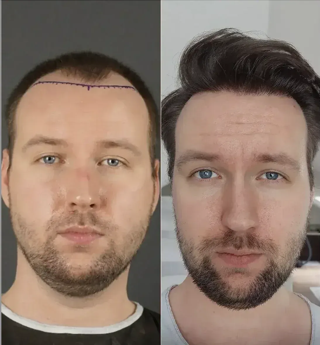
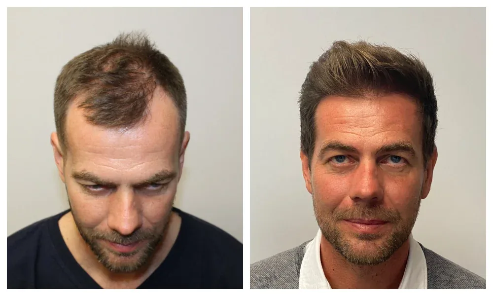
A study tells you something about groups. Only a doctor who has seen your photos can say what applies to your hair. The medical team at the clinic at the top of our ranking offers a free assessment.
Goes to the clinic at the top of our ranking.