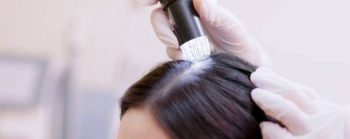
What is donor area trichoscopy, and what does it measure?
Trichoscopy means examining the scalp under magnification. It's what turns a donor assessment into something more than an eyeball estimate.
With trichoscopy, the scalp is examined under magnification and the donor assessment moves from an eyeball estimate to a concrete finding. The examination reveals miniaturisation, hairs of varying thickness and how the units are made up, things you cannot see with the naked eye.
What is trichoscopy?
Trichoscopy is dermatoscopy of the hair and scalp: the scalp is viewed through a magnifying instrument, either a small handheld device or a digital camera, usually under polarised light and often with a fixed-size field of view so the counts can be compared. The equipment is inexpensive and adds only a few minutes to the consultation.
What does trichoscopy show that you can't see with the naked eye?
It shows whether hairs vary in thickness, what share are thin and vellus hairs, how many hairs each unit has and how densely the units sit in a known area. It also reveals changes around the follicles and scarring where the openings have gone.
- Variable hair diameter. Adult hairs in the same small patch that clearly differ in thickness. This is the sign of androgen-driven miniaturisation, and the single most important finding when the donor area is assessed.
- Share of thin and vellus hairs. If the proportion of vellus hairs is rising, the follicles are shrinking.
- Hairs per follicular unit. How many hairs each cluster holds, from one up to four. This number converts grafts into hairs.
- Unit density. Counted in a patch of known size, not estimated.
- Changes around the follicles. Pigment or a brownish ring around each opening, linked to the inflammatory side of androgenetic alopecia.
- Signs of scarring. Openings that have disappeared where follicles used to be. This points to a scarring condition rather than ordinary hereditary hair loss.
Why does trichoscopy matter so much when the donor area is going to be used?
Because the back of the head can look dense even though many of its follicles are already shrinking, and grafts from such an area keep shrinking after they are moved. Xu and colleagues pointed out in 2026 that a spared occipital area is more often assumed than checked.
A patch can look completely normal to the eye even though many of its follicles are shrinking. A thin hair still takes up space and still reflects light. By the time you can see the thinning, it has been going on for a while.
This gap between how the back of the head looks and how things really are is the heart of the problem. A donor area that looks healthy but is quietly miniaturising produces grafts that carry on with the same process in their new location. At twelve months, the result looks good. In the years that follow it fades, and the reserve has already been used.
Xu and colleagues raised this in 2026. After comparing the crown and the back of the scalp in pairs, they argued that a spared occipital area is more often assumed than checked, and that donor-area hair loss can therefore be missed. Trichoscopy takes the guesswork out of it.
Where on the head should trichoscopy be done?
In several places, not just the middle of the back. A full examination covers the middle of the back of the head as a reference, the upper part towards the crown, the skin above and behind the ears, the nape hairline and the recipient area for comparison.
A single image from the middle of the back of the head is not an assessment. Density is not the same everywhere at the back, and neither is how stable the hair is. The edges usually thin first.
- The middle of the back of the head, as a reference.
- The upper part of the donor area, near the crown, where harvesting easily creeps too high.
- The skin around the ears, where thinning from below shows up first.
- The nape hairline. It is not part of the safe zone and is often thinned.
- The recipient area, for comparison. The difference in miniaturisation between the back and the top tells you a lot in itself.
What does good documentation of trichoscopy look like?
The findings are recorded as numbers, not just observations, so your donor area can be compared with itself in a few years. Without numbers, nobody can say for sure whether anything has changed. So ask whether the clinic keeps the images, whether you get a copy and whether the measurements are given as numbers, and consider taking standardised photos with the same hair length and lighting.
How do you know whether the clinic has actually assessed your donor area?
Whether the clinic owns a trichoscope is beside the point. Ask whether it was used on you, where on your head, and what it found. "Your donor area looks fine" is an impression, whereas "62 units per cm² at the middle of the back, 54 above the ears and 2.2 hairs per unit" is something you can build a plan on.
Sources
- Xu Y, et al. Case Report: Paired vertex-occipital assessment reveals donor-area involvement in diffuse unpatterned alopecia. Frontiers in Medicine, 2026;13:1797275. pubmed.ncbi.nlm.nih.gov/41982548
- Maas D, et al. Rethinking the occipital scalp as a control in advanced androgenetic alopecia. Journal of the American Academy of Dermatology, 2026 (ahead of print). pubmed.ncbi.nlm.nih.gov/42398778
- Jimenez F, Ruifernández JM. Distribution of human hair in follicular units. A mathematical model for estimating the donor size in follicular unit transplantation. Dermatologic Surgery, 1999;25(4):294-298. pubmed.ncbi.nlm.nih.gov/10417585
- Gupta AK, Venkataraman M, Talukder M, Bamimore MA. Finasteride for hair loss: a review. Journal of Dermatological Treatment, 2022;33(4):1938-1946. pubmed.ncbi.nlm.nih.gov/34291720
- Romera de Blas C, Vega Díez D, Ricart Vayá JM, Gómez Zubiaur A. Complications in follicular unit excision hair transplantation: current evidence and practical approaches. Frontiers in Medicine, 2026;13:1750989. pubmed.ncbi.nlm.nih.gov/41709896
This article is based on published research and standard clinical practice. It is general information, not medical advice, and it never takes priority over what your own surgical team tells you. If they say something different from what you read here, follow their advice.
Wondering what's possible for your hair?
Send three sharp photos of your hair, from the front, the top and the back. You'll get an honest answer on what can be done, roughly how many grafts it would take and what it costs.
- Free and with no obligation
- Photos are assessed by the clinic's medical team
- Replies usually within an hour in the daytime
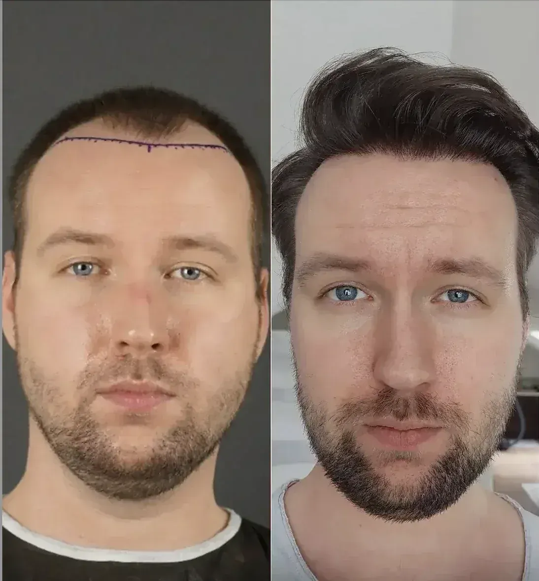
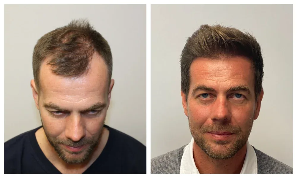
A study tells you something about groups. Only a doctor who has seen your photos can say what applies to your hair. The medical team at the clinic at the top of our ranking offers a free assessment.
Goes to the clinic at the top of our ranking.