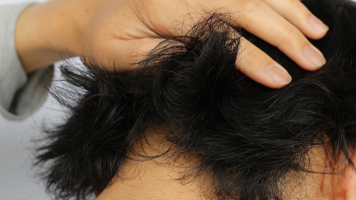
Do FUE dot scars show, and how long do they take to heal?
Every FUE extraction leaves a tiny, round scar, usually a shade paler than the surrounding skin. The result is final at months 6 to 12, and extraction density decides most of how much it shows.
Every graft taken with FUE leaves a small, round dot that usually ends up a shade paler than the surrounding skin. With normal density and a bit of length in the hair, nobody sees them. How visible they are depends on punch size, how densely the extractions are placed, colour contrast and how short you cut your hair.
What happens to the skin where each graft was taken?
The punch makes a small, round wound through the full thickness of the skin, and it heals without stitches. A scab forms within a few days, and over the following weeks the wound shrinks until new skin has closed it. In the end you are left with a small, round dot, usually a shade paler than the rest of the skin. Loss of colour in the dots is described as a complication in the literature, and they never disappear. So FUE is not a scarless method.
The punch makes a small, round hole through the full thickness of the skin in the donor area. The holes aren't stitched but are left to close on their own. For the first few days each one is covered by a tiny scab. Over the following weeks the hole shrinks and new skin grows over it.
What remains is a small, round dot. In most people it is a shade paler than the surrounding skin, because scar tissue has less pigment. Loss of colour at the extraction sites is described in the literature as a known donor area complication after FUE.
The dots stay for good. We say this plainly because FUE is sometimes sold as a "scarless" method. It is more accurate to say that one long scar has been swapped for many tiny ones.
How long does it take for the dot scars to heal fully?
Days 1 to 3: small, pink scabs, and the skin may be tender. Days 4 to 10: the scabs fall off once you start washing, but the pinkness stays. Weeks 2 to 6: the redness fades, more slowly in fair skin types. Months 2 to 4: the dots look roughly as they will, and any shedding recovers. Months 6 to 12: final result. Only now can the donor area be judged honestly.
- Days 1 to 3. Each extraction site gets a small scab. The skin is often pink and may feel tight or sore.
- Days 4 to 10. The scabs come away once you start washing again according to the clinic's plan. The pinkness remains.
- Weeks 2 to 6. The redness fades bit by bit. If you have fair skin, it usually takes longer.
- Months 2 to 4. The dots start to look the way they will end up. If you have had shedding in the donor area, it is usually on its way back.
- Months 6 to 12. Now you see the final result. Before this, it isn't fair to judge the donor area.
What decides whether the dot scars show at all?
Four things. Extraction density carries the most weight: a few extractions disappear in the hair, while many merge into a paler patch. The dot is as large as the punch. Pale skin and dark hair together reveal the dots first. And hair length decides a lot: with more than 6 mm of hair you won't see them, with a number one or a shaved head you will. If you stick to a number three or longer, they will probably never show.
Whether your donor area goes unnoticed or becomes something you want to hide comes down mostly to four factors.
- How densely the extractions are placed. This carries the most weight. If the dots are far apart, they blend into the hair. If they are close together, they merge into a visibly paler patch.
- Punch diameter. The dot ends up roughly the same size as the punch that made it.
- Colour contrast. Pale skin with dark hair above gives the greatest contrast and reveals the dots first. Lighter hair, or dark hair on darker skin, is more forgiving.
- How long your hair is. At 6 mm or more, a well-harvested donor area is usually impossible to tell apart from untouched skin. If you use a number one clipper or shave your head, you will see the dots in almost any light.
That last point is worth thinking through in advance. If you wear your head shaved, you will see the dots. If you stick to around a number three or longer, you will probably never notice them.
Which less common complications can occur as the donor area heals?
Besides the usual loss of colour, thickened scars, meaning raised, thick scar tissue, can form. This is uncommon and mostly depends on how you yourself scar. If you have had keloids or thick scars before, say so at the consultation, because it changes the risk with both FUE and FUT (strip method). Small cysts in the skin usually clear up on their own. Report them instead of squeezing them. Depletion of the donor area also belongs on this list.
How can visible dot scars be hidden?
The simplest and most effective option is to let your hair grow longer, and that is what most people do. Scalp micropigmentation adds tiny pigment dots that soften the contrast with the pale dots, and it is the most reliable measure for short hair. Grafts from the beard or body can be implanted into a depleted area, but this draws on your reserve. The scars stay. What you change is how visible they are.
- Let your hair grow. It is simple, it works best, and most people end up doing it.
- Scalp micropigmentation. Tiny dots of pigment make the pale dots stand out less against the skin. No method is more reliable against visible donor area scars, and it works well with short hair too.
- Grafts into the donor area. Beard or body hair is occasionally used to give a depleted area more density. Since you are using donor hair to fix a donor problem, the method has clear limits.
None of these solutions makes the scars go away. They make them less visible, and for most people that is exactly what counts.
Sources
- Romera de Blas C, Vega Díez D, Ricart Vayá JM, Gómez Zubiaur A. Complications in follicular unit excision hair transplantation: current evidence and practical approaches. Frontiers in Medicine, 2026;13:1750989. pubmed.ncbi.nlm.nih.gov/41709896
- Rassman WR, Bernstein RM, McClellan R, Jones R, Worton E, Uyttendaele H. Follicular unit extraction: minimally invasive surgery for hair transplantation. Dermatologic Surgery, 2002;28(8):720-728. pubmed.ncbi.nlm.nih.gov/12174065
- Chauhan K, Tandon M, Kumar A, Taneja N, Hamid SAT. A comprehensive review of evolution of advanced follicular unit excision systems. Journal of Cutaneous and Aesthetic Surgery, 2025;18(2):69-77. pubmed.ncbi.nlm.nih.gov/40212421
- Kim J, Ko YU, Yi KH. The condition of hair follicles produced by different punching methods during FUE surgery. Journal of Cosmetic Dermatology, 2024;23(12):4202-4207. pubmed.ncbi.nlm.nih.gov/39152658
This article summarises published research and common clinical practice. It is meant as general information and is not medical advice. Nor does it replace the advice you get from your own surgical team. If your team tells you something different from what is written here, follow your team.
Wondering what's possible for your hair?
Send three sharp photos of your hair, from the front, the top and the back. You'll get an honest answer on what can be done, roughly how many grafts it would take and what it costs.
- Free and with no obligation
- Photos are assessed by the clinic's medical team
- Replies usually within an hour in the daytime
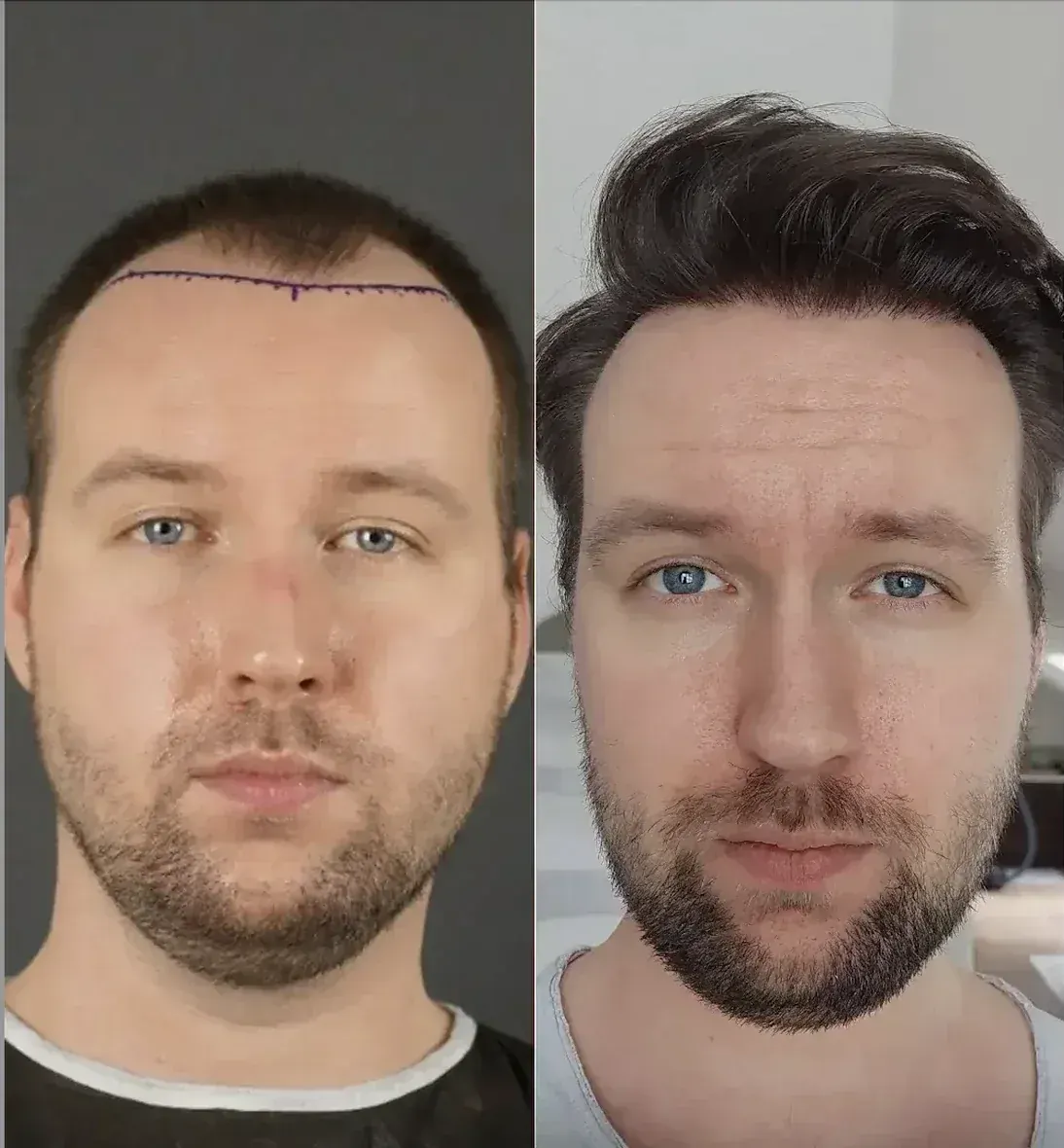
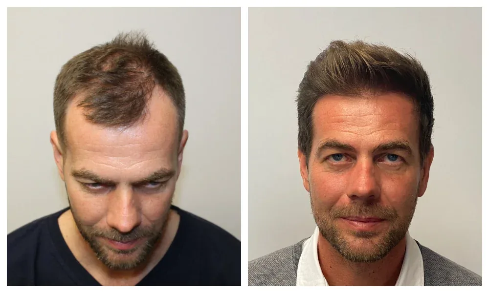
A study tells you something about groups. Only a doctor who has seen your photos can say what applies to your hair. The medical team at the clinic at the top of our ranking offers a free assessment.
Goes to the clinic at the top of our ranking.